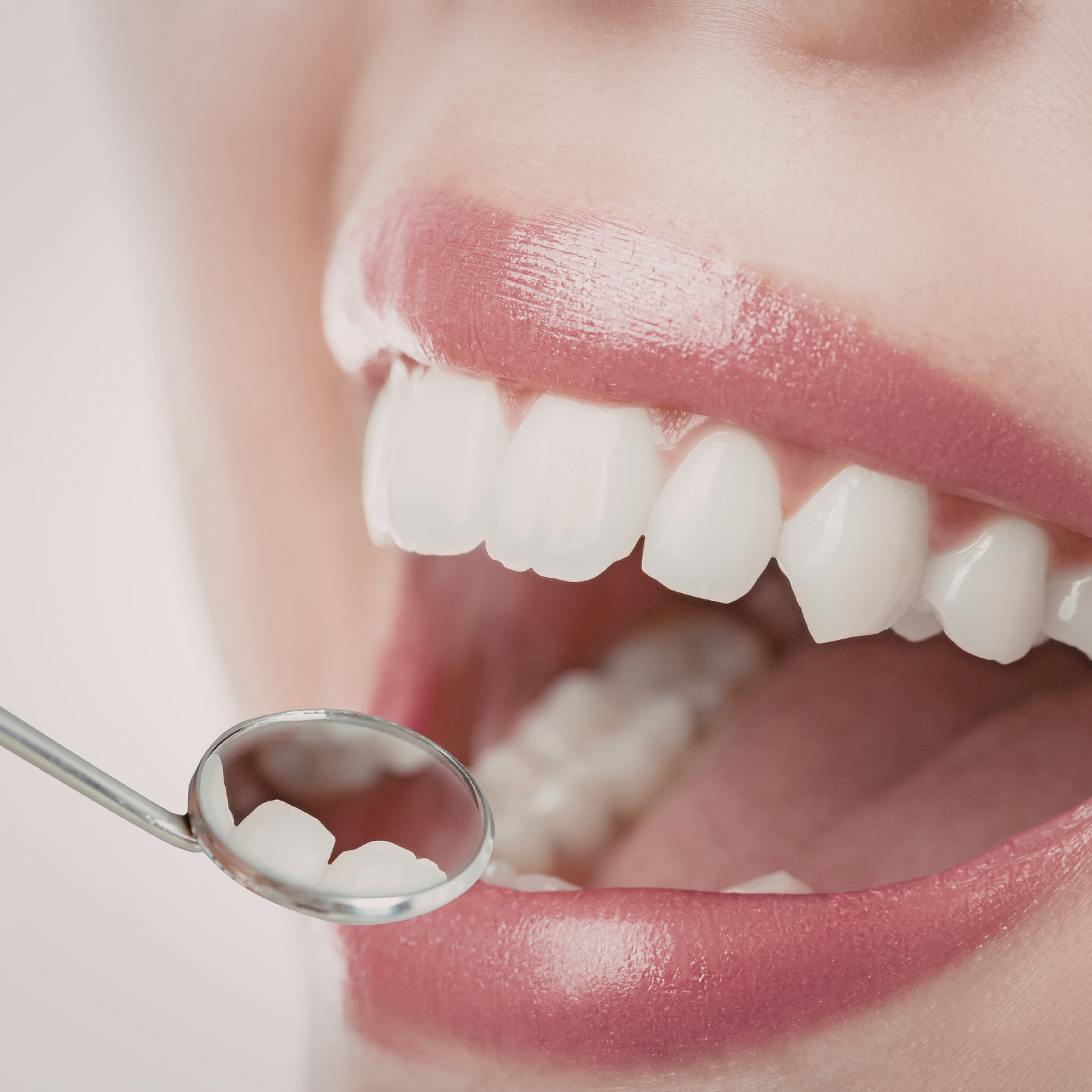
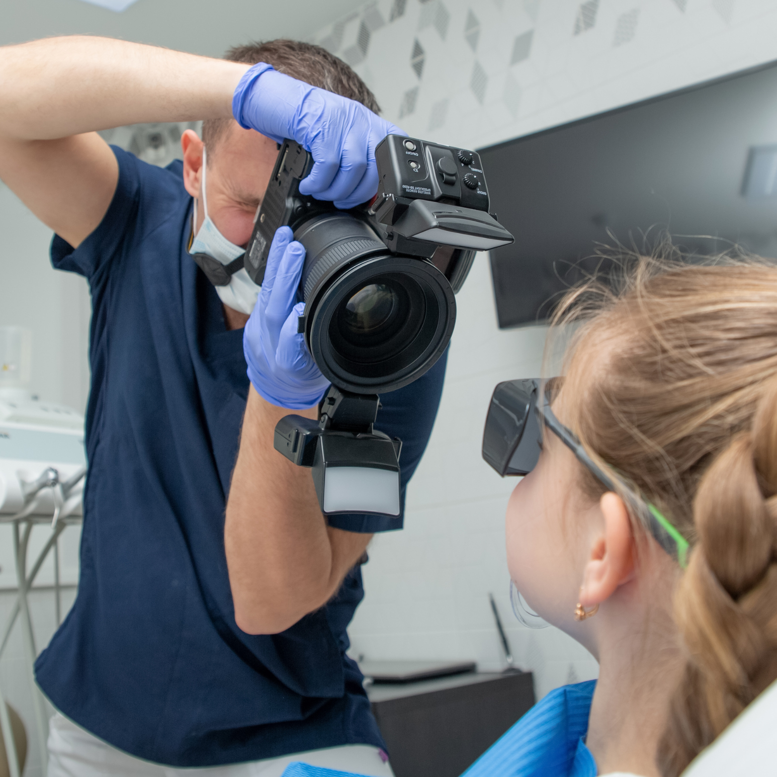
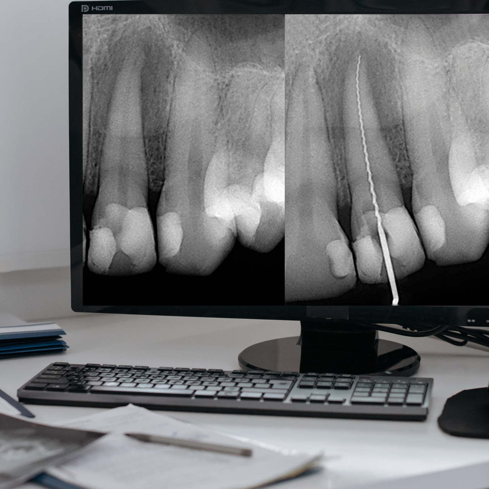
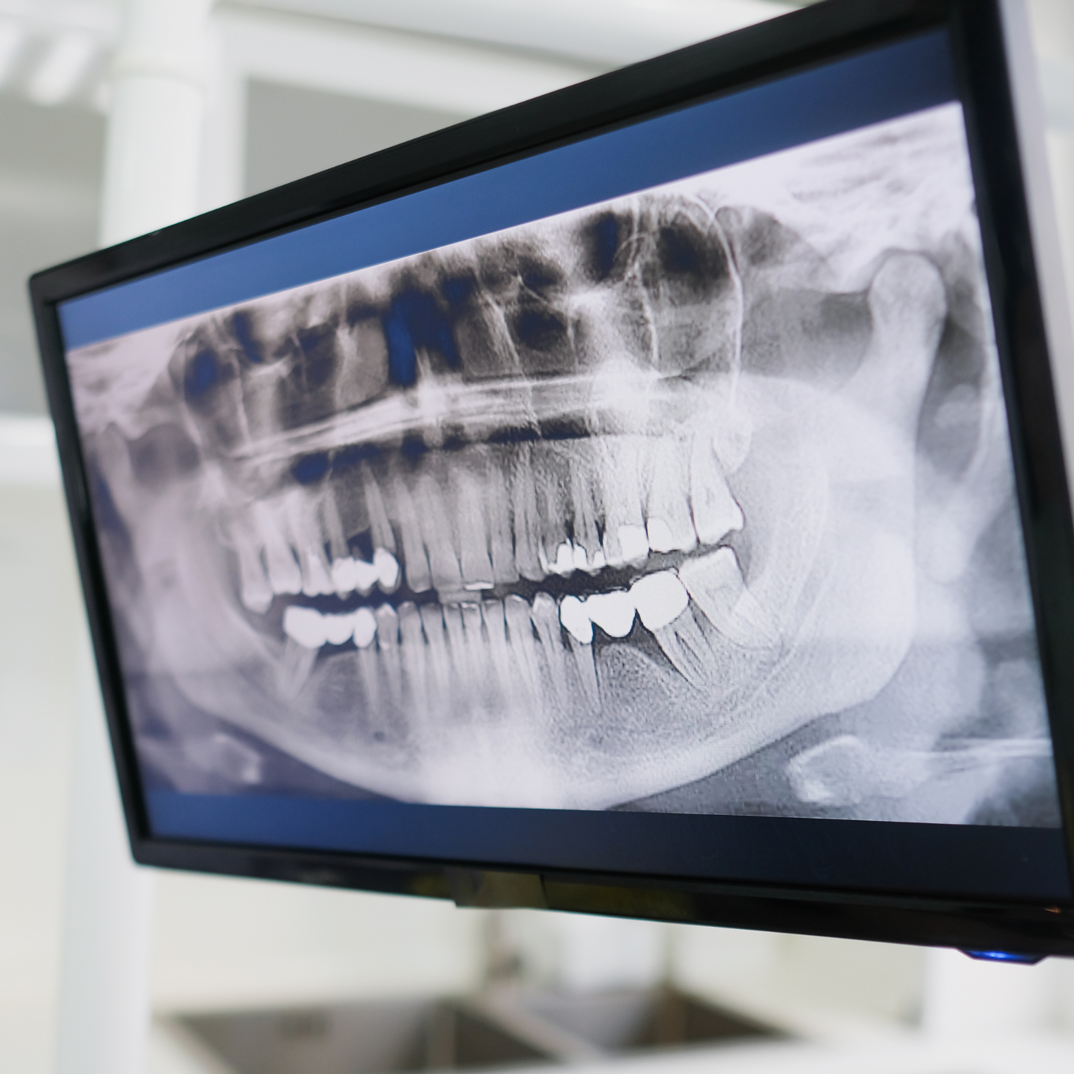
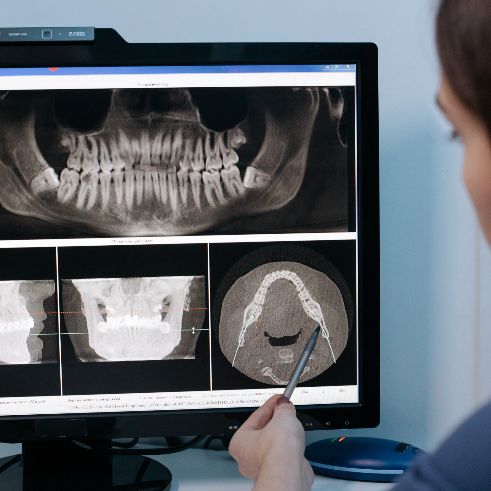
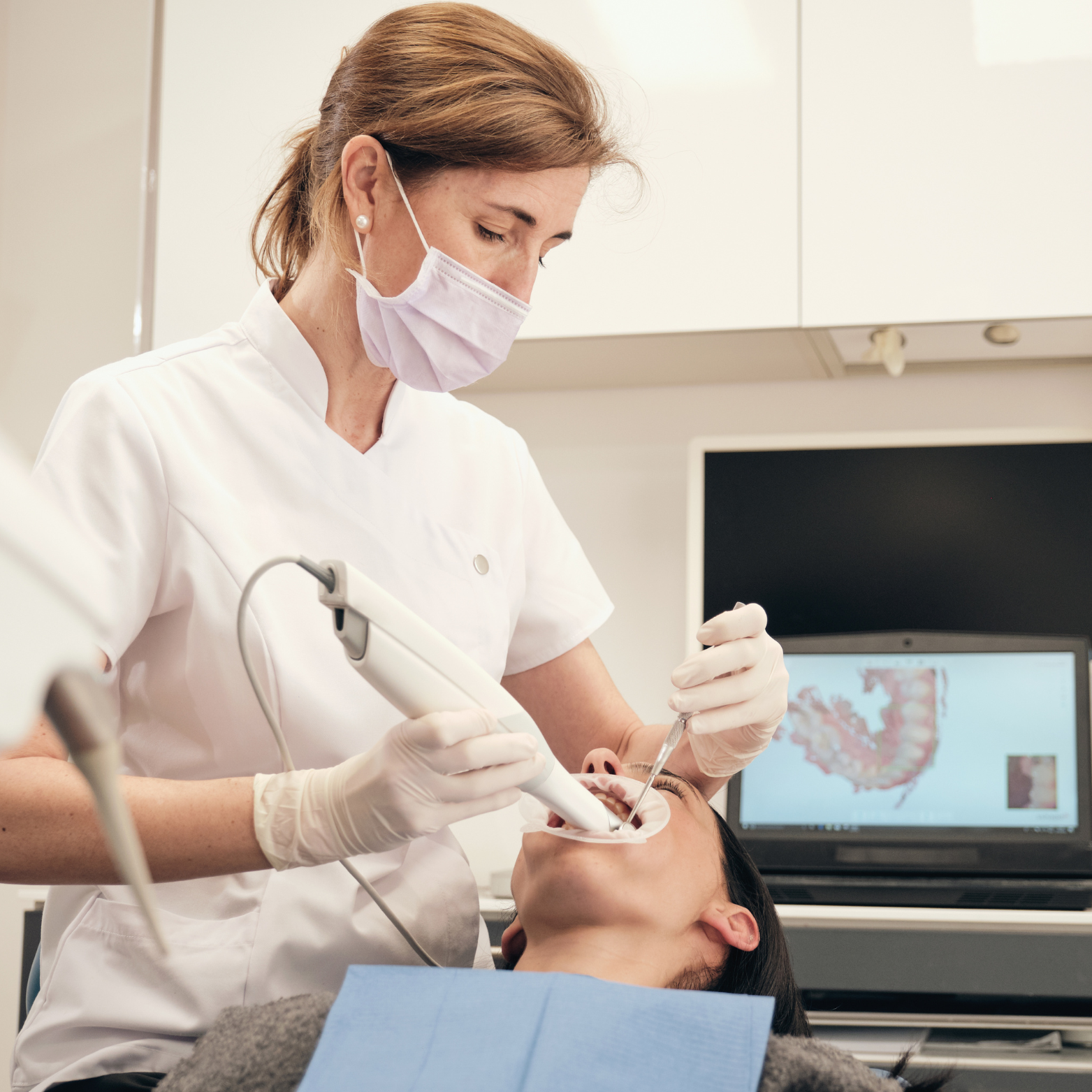
Dental Examination
-
DENTAL EXAMINATION
The right treatment begins with a thorough examination and accurate diagnosis.
Our comprehensive dentistry approach ensures you receive a clear, personalized treatment plan with options tailored to your condition and lifestyle. We explain each option in plain language so you can understand the benefits, risks, timeline, and costs, empowering you to choose the treatment that best enhances your comfort, function, and quality of life.
Because we believe no single plan fits everyone, our team collaborates with you to create a solution that respects your needs, priorities, and long-term goals.
-
DENTAL PHOTOGRAPHY
Dental photography is one of the most powerful tool we use to communicate with you—providing a clear, accurate visual record of your oral health so you can fully understand your condition before making any decisions.
High-quality photos let our team point out issues, compare progress, and illustrate treatment options in a way words alone cannot, giving you confidence and clarity about recommended care.
Because we believe that seeing the full picture empowers you to take an active role in your treatment, ask informed questions, and choose the path that’s right for you.
-
INTRAORAL X-RAY
High-quality Intraoral X-rays reveal the fine details beneath the gum-line, bone and tooth surfaces that the naked eye cannot detect, helping ensure accurate diagnoses and reducing the risk of missed important information.
With precise imaging, our clinicians can identify decay, bone loss, root issues, and hidden infections early, allowing for targeted treatment plans and greater peace of mind.
Your safety is our top priority: we adhere to strict radiation protection standards and use modern, low-dose equipment and shielding protocols to minimize exposure while delivering the diagnostic clarity you deserve.
-
EXTRAORAL X-RAY
We offer panoramic and cephalometric imaging to efficiently screen for bone and tooth lesions, delivering comprehensive views of the entire jaw, teeth, and surrounding structures in a single, low‑radiation scan. This advanced diagnostic tool helps our dentists detect abnormalities early, plan treatments more accurately, and monitor changes over time to ensure optimal oral health and predictable treatment outcomes.
Our extraoral X‑ray technology delivers exceptional image quality: combines advanced sensors and precise beam collimation to produce sharp, high‑resolution images that reveal fine anatomical detail for accurate diagnosis and treatment planning, while strict protocols and built‑in safety features keep radiation exposure to an absolute minimum so you can feel secure about radiation hazards.
-
CBCT (3D X-RAY)
3D imaging in dental X‑ray is essential for complex cases such as root canal treatments, implant placements, and others oral surgeries because it reveals detailed views of the tooth, bone, and surrounding structures that conventional 2D images can miss. By visualizing canal anatomy, bone density, proximity to nerves and sinuses, and the true extent of pathology, clinicians can plan procedures more precisely, anticipate potential complications, choose the most appropriate instruments and approaches.
We believe complex cases demand greater detail to support confident decision‑making, reduce intra-operative surprises, and achieve safer, more predictable outcomes with fewer complications.
-
INTRAORAL SCANNER
Intraoral scanners propel us into the digital world by capturing high-quality, precise 3D images of your teeth and oral structures quickly and effortlessly; their speed reduces chair time, their accuracy improves restorative fit and treatment planning, and their ease of use enhances patient comfort and communication, allowing our team to deliver faster, more predictable dental care with digital workflows that streamline impressions, labs, and record-keeping.